MRI vs MRA - What needs considering?
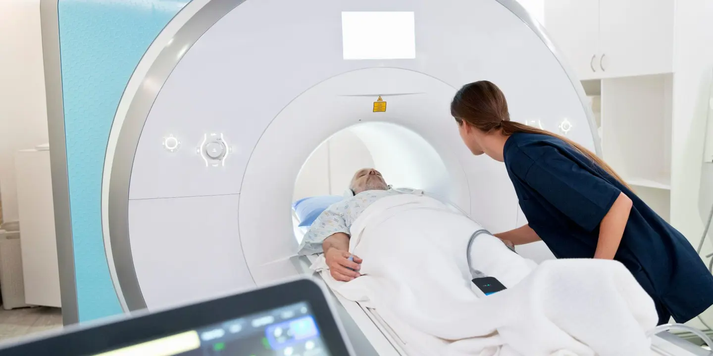
Let’s take the second of these factors first. When it comes to facilities, if you have access to a 3T scanner then, often, a plain MRI Scan can answer the question. The detail that can be achieved with a 3T scanner is superior to a 1.5T scanner and therefore allows the subtle findings to be identified more readily. It must, however, be noted that this is not always the case. Even with a 3T scanner it may be necessary to perform an arthrogram.
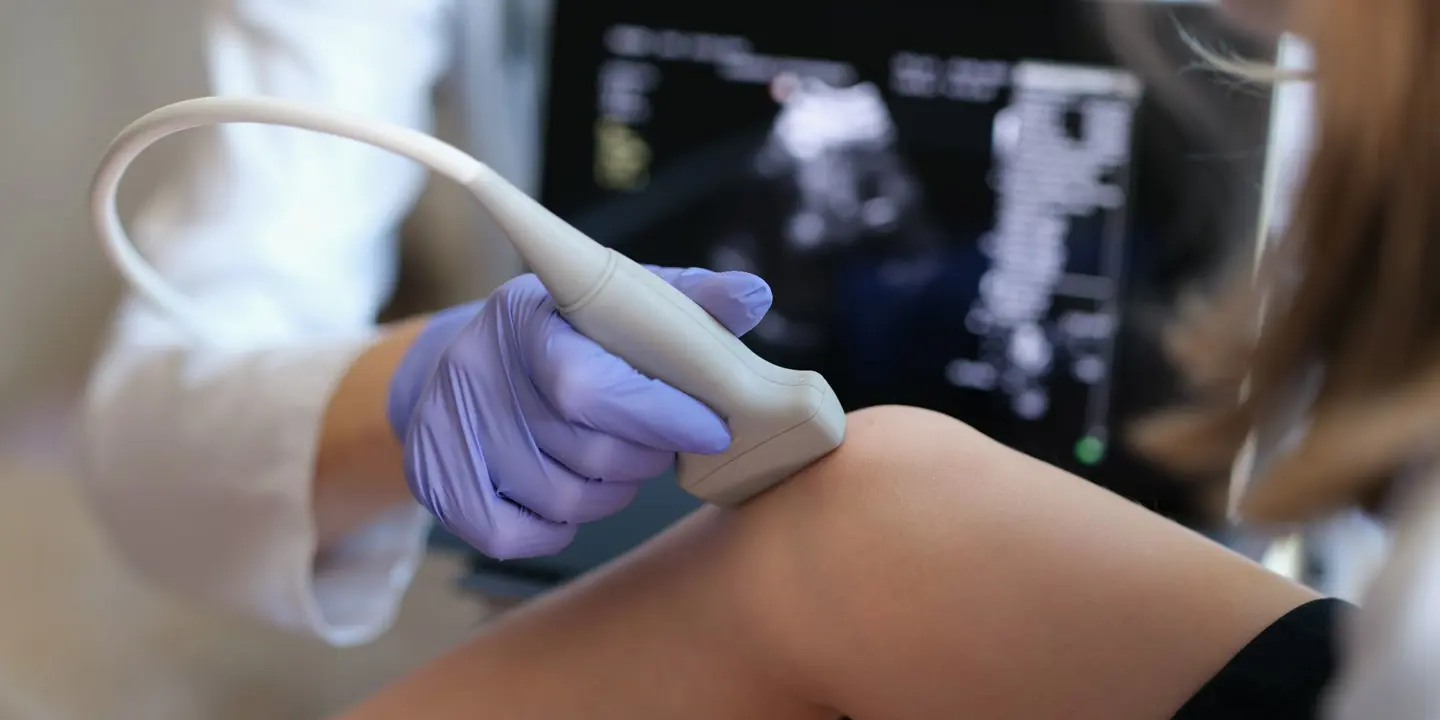
When performing an arthrogram, it is necessary to inject a contrast agent directly into the joint in question, so therefore the examination takes on a 2-stage process. Stage 1 is to perform the injection and stage 2 is to perform the MRI Scan. For this, it is essential to have the Radiologist present, who can perform the injection with either ultrasound or fluoroscopic guidance. This makes the procedure highly operator dependent.
Time can also be a factor. In the wrist, for example, it would be ideal to do a 3-compartment arthrogram, with an injection being performed in between each scan. However, the whole procedure in this case would take more than 2 hours, which is impractical. Therefore it is general practice to perform a uni-compartment wrist arthrogram, which in most cases is the radiocarpal compartment.
All this aside, the single most important factor when deciding between a MRI Scan or an MR arthrogram is the clinical question that needs to be answered. Let’s examine the 2 most common arthrographic studies; Shoulder and Hip.
With the shoulder, if the clinical question relates to rotator cuff pathology or AC joint arthropathy, then MRI alone should suffice. One could argue that Ultrasound would be the investigation of choice, but in any case there is no need for an arthrogram. If intra-articular pathology is suspected, for example in instability such as labral injuries, ligamentous injuries or cartilage abnormalities, then an MR arthrogram would be considerably more sensitive in this regard.
With the hip joint, it is a similar decision-making process. For infra-articular pathology regarding labral pathology and cartilage abnormalities, one should consider an arthrogram. For superficial conditions such as trochanteric bursitis, an MRI or even Ultrasound will be the investigation of choice.
We all know that patients often present in a complex manner, and it may be difficult to pin-point, pre-test, what the pathology is likely to be. In that case, it could be challenging to decide how to proceed, but a pragmatic and non-invasive way could be to perform a plain MRI Scan and, if that doesn’t provide the diagnostic answer, then one could proceed to an arthrogram at a later stage.
The other main joint that we asked to perform arthrograms on is the wrist joint. This, again, depends on the clinical question to be answered. If a scaphoid fracture union needs to be assessed, this can only be done reliably by performing an arthrogram, which could be CT or MR based. A ligamentous injury may be diagnosed on a plain MRI Scan, but the sensitivity is greatly increased with an MR arthrogram.
I have also occasionally been asked to perform an arthrogram of the elbow, and this can be useful when assessing a pre-surgical patient.
Personally, I have never been asked to perform an arthrogram on any other joint, but I know of centres that perform ankle arthrograms to assess for ligamentous injuries.
In summary, in the correct clinical context, MR arthrography is superior than a plain MR Scan. 3T scanning has brought the two techniques closer together. The clinical examination and pre-test query remains vital in determining whether to subject patients to what is, after all, an invasive test with a 2-stage process.
Finally, in cases where there is genuine uncertainty, it can be useful to have a discussion with your MSK Radiologist to decide the best course to take.
Dr Qaiser Malik MRCP FRCR
Consultant MSK Radiologist
Vista Health


